biography
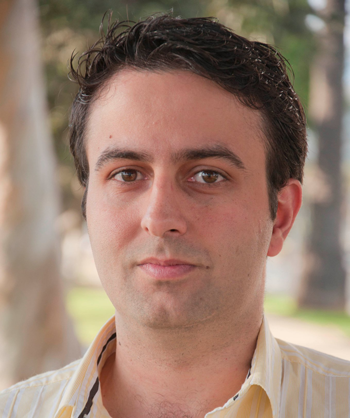
Dr. Hojjat is an Assistant professor at University of Toronto and an affiliate scientist under the Hurvitz Brain Sciences program at Sunnybrook Research Institute. His research involves developing multimodal/ functional imaging applications to assess degenerative diseases and neurological disorders. His current research focus involves utilization of perfusion imaging modalities in two distinct applications: 1) to regionally quantify effect of collaterals on tissue salvage after stroke, 2) to quantify the global and regional perfusion at different stages of multiple sclerosis and to analyze the implication of these measures in cognition. Previously, he worked as a research scientist at GE Healthcare at St. Joseph hospital in London, ON. where he actively collaborated with the University of Western Ontario, Robarts Research Institute, Lawson Health Research Institute and London Health Sciences Centre. His research included automated multimodal image-based techniques for quantitative evaluation of spinal disease and disorders as well as treatment effect. During his PhD, he developed an automated multi-modal µCT, µMR based tool for analysis of tumour burden and mechanical stability in metastatic spine. Throughout his career, Dr. Hojjat has co-invented 2 patents, co-authored 14 full papers and presented his research through over 20 peer reviewed conference papers.
Area of Interest
1) Multimodal medical image analysis
2) Perfusion and permeability
3) Multimodal image registration
4) Software and hardware development for biomedical engineering applications
top publication
1. Hojjat SP, Cantrell CG, Vitorino RC, Feinstein A, Shirzadi Z, MacIntosh B, Crane D, Zhang L, Morrow S, Lee L, O’Connor P, Carroll TJ, Aviv R (2015). Regional reduction in cortical blood flow among cognitively impaired adults with relapsing-remitting multiple sclerosis patients. Multiple Sclerosis Journal (In press).
2. Hojjat SP, Kincal M, Vitorino RC, Cantrell CG, Feinstein A, Shirzadi Z, MacIntosh B, Crane D, Zhang L, Morrow S, Lee L, O’Connor P, Carroll TJ, Aviv R (2015) Cortical perfusion alteration in normal appearing gray matter is most sensitive to disease progression in relapsing remitting Multiple Sclerosis. American Journal of Neuroradiology (In press).
3. Vitorino RC, Hojjat SP, Cantrell CG, Feinstein A, Lee L, O’Connor P, Carroll TJ, Aviv R (2015) Regional Frontal Perfusion Deficits in Relapsing Remitting Multiple Sclerosis – a Marker of Disease Severity? Multiple Sclerosis Journal (In press).
4. Hojjat SP, Cantrell CG, Carroll TJ, Vitorino RC, Feinstein A, Zhang L, Symons SP, Morrow S, Lee L, O’Connor P, Aviv R (2015). Perfusion reduction in the absence of structural differences in cognitively impaired versus unimpaired RRMS patients. Multiple Sclerosis Journal (In press).
5. Hojjat SP, Beek M, Akens MK, Whyne CM (2011) Can micro-imaging based analysis methods quantify structural integrity of rat vertebrae with and without metastatic involvement?, J Biomech. 2012 Aug 1. [Epub ahead of print].
6. Hojjat SP, Foltz W, Wise-Milestone L, Whyne CM. (2011) Muti-modal µCT /µMR based Semi-Automated Segmentation of Rat Vertebra Affected by Mixed Osteolytic / Osteoblastic Metastases, Med Phys. 2012 May; 39(5):2848-53.
7. Absolute quantification of normal appearing and lesional tissue in RRMS. Hojjat SP, Kincal M, Vitorino RC,Cantrell CG, Feinstein A, Shirzadi Z, MacIntosh B, Crane D, Zhang L, Morrow S, Lee L, O’Connor P, Carroll TJ,Aviv R. American Society of Neuroradiology (ASNR) 2016, Washington DC, USA.
8. Comparison of quantitative cerebral blood flow between Bookend and pseudo-Continuous Arterial Spin Labelling in Relapsing Remitting Multiple Sclerosis. Hojjat SP, D’Ortenzio R, Vitorino RC, Cantrell CG, Feinstein A, Lee L,O’Connor P, Carroll TJ, Aviv R. ASNR 2016, Washington DC, USA.
9. Effect of collaterals on clinical presentation, baseline imaging, complications and outcome. Fanou F, Knight J, Aviv R, Hojjat SP, Symons S, Zhang L, Wintermark M, Radiology Society of North America (RSNA), 2014.
10. Automatic Data Driven Labeling of Lumbar Spine Structures in MRI. Ben Ayed A., Hojjat SP, Punithakumar K,Garvin G., RSNA 2014.
11. Automated 3D visualization and assessment of the inter-vertebral disc displacements in spine imaging. Ben Ayed A., Punithakumar K, Hojjat SP, Garvin G., Accepted for oral presentation at Computer Assisted Radiology and Surgery conference, Heidelberg, Germany. June 26th – June 29th, 2013.

