Neurocysticercosis in a Native Caribbean Woman Living in New York
Corey Bascone1, Mutafa S. Mawih2, Uri Goldberg2, Mukhamad S. Valid 2*
Affiliation
- 1Medical student, American University of Antigua (AUA) College of Medicine
- 2Clinical resident, Kingsbrook Jewish Medical Center
Corresponding Author
Mukhamad S. Valid, Clinical resident, Kingsbrook Jewish Medical Center, New York, USA. E-mail: mswalid@yahoo.com
Citation
Valid, M. S. et al. Neurocysticercosis in a Native Caribbean Woman Living in New York. (2015) Int J Neurol Brain Disord 2(1): 1- 3.
Copy rights
© 2015 Valid, M. S. This is an Open access article distributed under the terms of Creative Commons Attribution 4.0 International License.
Keywords
Neurocysticercosis; Seizure; Caribbean
Abstract
We report a case of neurocysticercosis in a 48-year old female who presented to the ED of a Brooklyn community hospital with a new-onset seizure and a recent travel history to the Caribbean island of Trinidad. Considering neurocysticercosis in the differential diagnosis of any adult presenting with a new onset seizure and/or neurologic manifestations is essential, especially among immigrants
Introduction
Cysticercosis is a parasitic tissue infection caused by larva of the tapeworm, Taenia solium. These larval organisms invade the muscles, the brain, and various other tissues; where they form cysts known as cysticerci. When these cysts form in the muscles, they are usually asymptomatic. Symptoms following brain involvement, exclusively referred to as neurocysticercosis, depend on the amount of damage caused to the central nervous system[1]. It is important to understand that the pathophysiology of this tissue infection is indeed different from Taeniasis, the intestinal infection. Taeniasis infection idevelops from ingesting larval-cyst-containing pork or beef meat, which is undercooked. The adult worm lives in the intestine of humans and only when humans shed the proglottids (containing eggs) in their feces and swine ingests it can cysticerci develop in the muscle of these livestock. In order to develop cysticercosis in humans, one must ingest feces from another infected human, or already have taeniasis and auto-infect themselves[2].
Cysticercosis is endemic in many regions around the world including areas of Central and South America, sub-Saharan Africa, India, and Asia[3-8]. Infection is found most often in the rural areas of these regions, where pigs are allowed to roam freely and are able to come in contact with human feces. However, cysticercosis itself is not considered to be common in the Caribbean[9], with the exception of Haiti, where several case reports have been published in the past decades[10,11]. Here we report a case of neurocysticercosis in a 48-year old female who presented to the ED of a Brooklyn community hospital in May 2014 with a new-onset seizure and a recent travel history to the Caribbean island of Trinidad.
Case Report
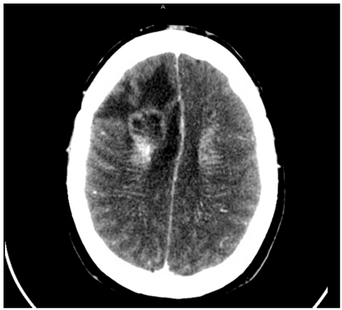
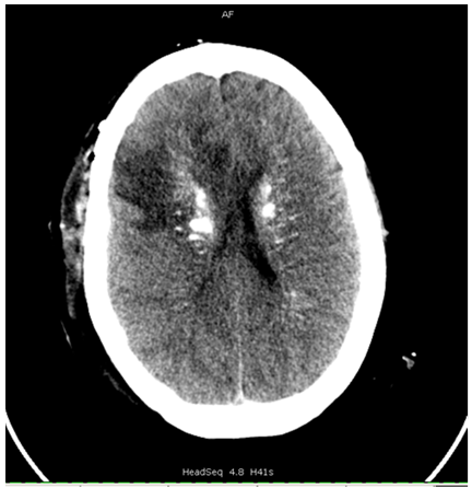
A previously well, 48-year old woman presented to the emergency department of Kingsbrook Jewish Medical Center (KJMC) in Brooklyn New York with a two-day history of headache of increasing intensity and one episode of generalized seizure. Upon arrival, the patient was alert, oriented to time, person and place and deemed a reliable historian based on her alertness and ability to provide a thorough history. With no significant past medical history, the patient denied the use of any over-the-counter or herbal medications, the use of illicit drugs, as well as any family history of seizure disorder. The patient further explained that her family was from Trinidad, that they were all healthy, and how she visited them quite often up until recent time. Her last travel to Trinidad was two years prior to that presentation and she denied any complaints after the last visit. Results of the physical exam showed no focal neurological signs. Upon brain imaging, CT scan of the head revealed multiple areas of calcifications throughout both right and left cerebral hemispheres, the basal ganglia, and the cerebellar nuclei. A localized contrast-enhancing lesion was also identified in the right frontal lobe, accompanied with a 0.3 cm midline shift and adjacent neurogenic edema [Figure 1A]. Following these findings, the patient agreed to undergo neurosurgery, where craniotomy and debulking of the lesion was performed [Figure 1B]. Tissue specimens were obtained during the operation from three different sites and sent for pathological examination. The first tissue specimen submitted for fresh frozen section was removed from the right frontal brain mass, consisted of grayish-white soft tissue, and measured in at 0.8 x 0.8 x 0.2 cm. A second specimen was surgically removed from another brain mass in the cerebral hemisphere, measured 1.5 x 1 x 0.3 cm, and consisted of whitish tissue fragments. The third and last tissue specimen submitted for frozen section was excised from the only other surgically accessible brain lesion, measured 1.5 x 1.5 x 0.5 cm and consisted of white friable and soft tissues.
Figure 1A: CT scan of the head on presentation showing Right frontal peripherally enhancing centrally necrotic lesion with slight right-to-left subfalcine herniation approximately 0.3 cm.
Figure 1B: CT scan of the head Status post debulking procedure(1st postoperative day)right frontal lobe lesion with routine postoperative appearance
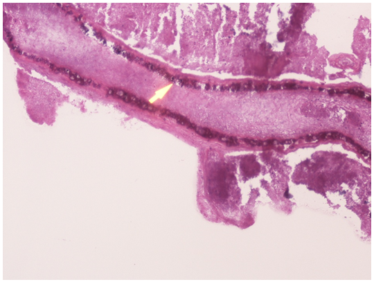
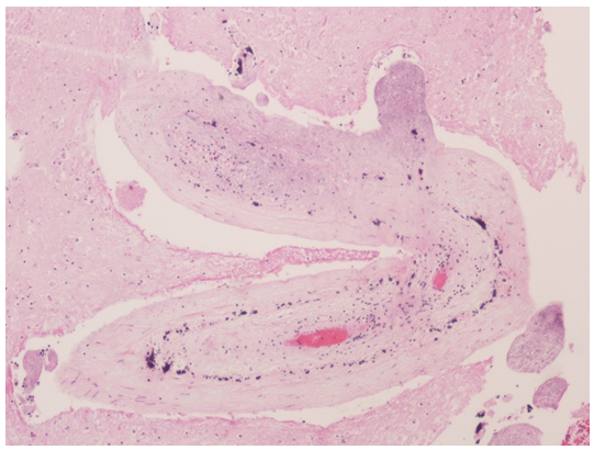
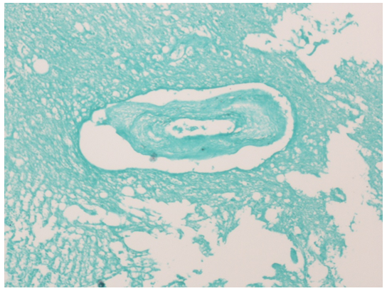
The pathologic report later revealed degenerating glial tissue, tubular areas with marked wall calcification, multinucleated giant cells, psammoma-like bodies and various foci of mononuclear cells [Figure 2A-C]. No evidence of malignancy was reported amongst these pathological findings and thus, the differential diagnosis of vascular malformation with marked wall calcification versus parasitic infection with cysticercosis was considered. Medical treatment was started empirically with albendazole, oral steroids and anticonvulsant therapy. The patient was discharged after thorough explanation of the findings and management of the suspected infection was conducted. Following the initiation of treatment, the patient remained free of clinical symptoms and serial CT scans of the brain displayed significant healing of the frontal lobe lesion at both the one and two month mark post-operatively; confirming the diagnosis of neurocysticercosis [Figure 3].
Figure 2A: Pathology slides showing degenerated neural tissue with tubular areas with marked calcification in the wall (arrow head) and lumen showing clear pale material.
Figure 2B: Cross-section of a hematoxylin & eosin stained tubular structure with marked wall calcification.
Figure 2C: Cross-section of the tubular structures with adjacent degenerative neural tissue in Gomori's Methenamine silver stain.
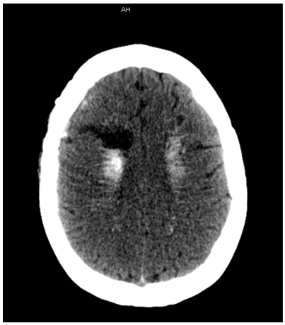
Figure 3: CT scan of the head 1 month after surgery showing near complete treatment response with decreased enhancement and decreased vasogenic edema surrounding a right frontal cavity representing a postsurgical site.
The patient completed her medication regimen, as well as physical therapy and six months later on January 6th 2015, the patient presented to the KJMC emergency department following a tonic-clonic seizure of 2-3 minute duration. The patient experienced the seizure while eating lunch at her place of work, did not recall what happened during the episode, and was disoriented to time and place for ten minutes following the seizure. At time of exam, no neurological symptoms were reported and CT scan of the head was negative for acute changes. While waiting for the results of the MRI, the patient was placed on 1000 mg of IV Dilantin. MRI later reported no new enhancements or lesions and focal gliosis of the post-operative cavity from prior craniotomy. With satisfactory imaging reports and no observable neurologic defects, it was determined that the patient should remain on an anti-epileptic for at least two years in order to prevent any additional seizures and ensure proper recovery.
Discussion
The clinical presentation of neurocysticercosis is quite variable, ranging from vague neurological symptoms, all the way to sudden death; with abnormal physical exam findings occurring in only 20% or less of patients[12]. Other signs and symptoms of neurocysticercosis include epilepsy, which is the most common symptom, appearing in over 70% of infected patients[13], as well as headache, dizziness, stroke, and neuropsychiatric symptoms. The appearances of these clinical findings depend on many factors including the location and size of the cystic space. The stages of neurocysticercosis vary depending on the viability of the larva involved and the immune response displayed by the host[14]. While still viable, the development of larval cysts are associated with minimal inflammation, also known as the vesicular stage. Once the worm dies after 2-6 years of infection, the cysticerci lose the ability to control the host's immune response, allowing the cyst wall to become surrounded and infiltrated by mononuclear cells. Inflammatory cells then enter the cyst fluid and this is known as the colloid stage. As the host immune response progresses, fibrosis encompasses the cysticerci, with consequential collapse of the cyst cavity, which is known as the granular-nodular stage. The dead parasite eventually decays into eosinophilic material and desiccates, leaving behind a calcified nodule. This final stage is known as the calcific stage as a result of the necrotic larva undergoing dystrophic calcification[15].
If the infection is found in one of the latter stages, as it was in our case, it is important to understand procedural precautions. For example, the larger the cyst cavity is, the more likely to develop edema, hydrocephalus, and displacement of brain structures; often making lumbar puncture with CSF analysis contraindicated. CT scanning or MRI, after the intravenous administration of contrast material is the imaging test of choice. If an eccentric scolex is seen within the cyst, neurocysticercosis may be diagnosed confidently. However, in the absence of neuroimaging, Enzyme-linked immune transfer blotting (EITB) is the most accurate and most practical serologic test used for screening, with a sensitivity of 90-100%[16]. This serologic screening tool should always be considered in a patient with a history of seizure and/or unexplained focal neurologic defects.
In a review of literature, a prospective study of 1800 seizure patients was noted. In that study, 1800 patients, who presented with seizures to 11 different U.S. emergency departments, were documented over a two-year period. Neurocysticercosis was found to be the etiologic agent in about 2 percent of these cases[17]. It was noted that the infection was observed at a much higher frequency amongst emergency departments in Los Angeles, Phoenix, and Albuquerque (5.7%), which happened to have a higher proportion of immigrant hispanic patients than the other hospitals included in the study[17]. Therefore, making the consideration of neurocysticercosis essential in the differential diagnosis of any adult presenting with a new onset seizure and/or neurologic manifestations, especially among immigrants. However, the possibility of diagnosis should not be limited to these regions, as in our case patient traveled to a non-endemic region, Trinidad. The case itself indicates that other risk factors associated with acquiring this disease are equally as important when exposure to infected carriers and contacts are considered[18–20]. To further defend this theory, a similar circumstance was reported in an Orthodox Jewish community in New York City. Although eating pork is not practiced among the community for religious reasons[21], neurocysticercosis was found amongst the population in patients suffering from seizures and accompanied radiologic evidence of cystic brain lesions.
The variety of clinical, radiological and epidemiological presentations of neurocysticercosis necessitate a high index of suspicion to be practiced by health care professionals in the present day. Furthermore, it is imperative that infectious etiology in general is considered in any previously well patient, presenting with newly formed neurologic symptoms and/or episodes of seizure. Maintaining high awareness of the various clinical scenarios possible for presentation, as well as further study of the disease is required in order to control the increase of neurocysticercosis incidence in the United States[12].
References
- 1. Johnson, R. T., Davis, L. E. Neurocysticercosis: Clinical Summary. (1993) Neurology Medlink.
- 2. Mandell, G. L., Bennett, J. E., Dolin, R. Principles and Practice of Infectious Diseases. 7thEdn. (2009) Elsevier Churchill Livingstone.
- 3. Montano, S. M., Villaran, M. V., Ylquimiche, L., et al. Neurocysticercosis: association between seizures, serology, and brain CT in rural Peru. (2005) Neurology 65(2): 229- 233.
- 4. Nicoletti, A., Bartoloni, A., Sofia, V., et al. Epilepsy and neurocysticercosis in rural Bolivia: a population-based survey. (2005) Epilepsia 46(7):1127- 1132.
- 5. Medina, M. T., Durón, R. M., Martínez, L., et al. Prevalence, incidence, and etiology of epilepsies in rural Honduras: the Salamá Study. (2005) Epilepsia 46:124-131.
- 6. Rajshekhar, V., Raghava, M. V., Prabhakaran, V., et al. Active epilepsy as an index of burden of neurocysticercosis in Vellore district, India. (2006) Neurology 67(12): 2135- 2139.
- 7. Ndimubanzi, P. C., Carabin, H., Budke, C. M., et al. A systematic review of the frequency of neurocyticercosis with a focus on people with epilepsy. (2010)PLoSNegl Trop Dis 4(11): e870.
- 8. Willingham, A. L.3rd., Engels, D. Control of Taeniasoliumcysticercosis/taeniosis. (2006) AdvParasitol 61:509-566.
- 9. Hotez, P. J., Bottazzi, M. E., Franco-Paredes, C., et al. The neglected tropical diseases of Latin America and the Caribbean: a review of disease burden and distribution and a roadmap for control and elimination. (2008) PLoSNegl Trop Dis 2(9): e300.
- 10. Case records of the Massachusetts General Hospital. Weekly clinicopathological exercises. Case 48-1984. A 43-year-old Haitian man with headaches and visual abnormalities. (1984) N Engl J Med 311(22): 1425- 1432.
- 11. Fleet WF 3rd., Morgan HJ., Lile S. Cystic brain lesions in a Haitian man. (1990) J Tenn Med Assoc 83(8): 400–401, 403.
- 12. Moskowitz J., Mendelsohn, G. Neurocysticercosis. (2010) Arch Pathol Lab Med 134(10):1560- 1563.
- 13. Chaoshuang, L., Zhixin, Z., Xiaohong, W., et al. Clinical analysis of 52 cases of neurocysticercosis. (2008) Trop Doct 38(3):192- 194.
- 14. Pearson, R. D.TaeniaSolium (Pork Tapeworm) Infection and Cysticercosis.
- 15. White, A. C. Jr. Neurocysticercosis: updates on epidemiology, pathogenesis, diagnosis, and management. (2000) Annu Rev Med 51: 187- 206.
- 16. Parasites – Cysticercosis. Center for Disease Control and Prevention(CDC).
- 17. Ong S, Talan DA, Moran GJ, et al. Neurocysticercosis in radiographically imaged seizure patients in U.S. emergency departments. (2002) Emerg Infect Dis 8(6): 608- 613.
- 18. Sorvillo, F. J., DeGiorgio, C., Waterman, S. H. Deaths from cysticercosis, United States. (2007) Emerg Infect Dis 13(2): 230- 235.
- 19. Earnest, M. P, Reller, L. B., Filley, C. M., et al. Neurocysticercosis in the United States: 35 cases and a review. (1987) Rev Infect Dis 9(5): 961- 979.
- 20. del la Garza, Y., Graviss, E. A., Daver, N. G., et al. Epidemiology of neurocysticercosis in Houston, Texas. (2005) Am J Trop Med Hyg 73(4): 766- 770.


